What is a dental X-ray?
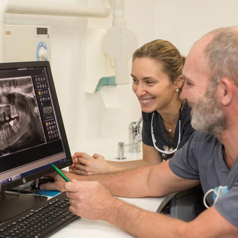
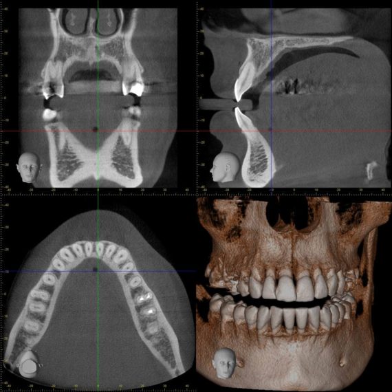
Radiology is an imaging technique based on the transmission of X-rays through the structures under examination to a sensor. The different densities of the tissues through which the X-rays pass induce variations in the quantity reaching the sensor. Anatomical structures then appear on the radiograph in grayscale.

Why are dental X-rays necessary?
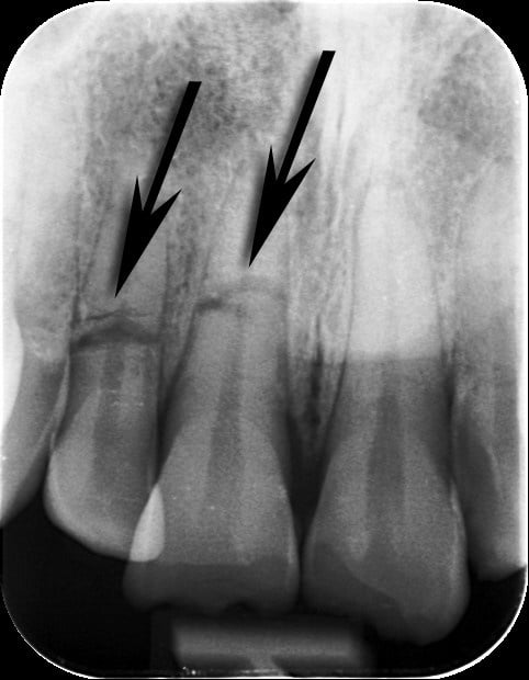
Dental radiographs represent a (currently) irreplaceable diagnostic tool and contribute to the planning of any treatment: detection of caries, periapical damage, bone lesions, periodontal disease with bone involvement, trauma assessment,…
Not all of these structures can be satisfactorily examined with the naked eye, and require X-rays.

What precautions should I take for dental X-rays?
X-rays are ionizing radiation and can have harmful effects on health in the event of long and repeated exposure and/or for high intensities.
Several scientific studies reported in the media point to a possible link between dental X-rays and the risk of cancer (meningioma). All these studies have major weaknesses and are based on obsolete radiology technology: analog radiology. Nevertheless, a reasoned use of X-rays in the dental field is essential.

Radiological procedures in dentistry (mainly intra-oral photography) expose patients to very little radiation. And technological developments with the advent of digital radiography have made it possible to considerably reduce radiation (50 to 75%).
All our dentists are licensed by the Swiss Federal Office of Public Health (FOPH) to carry out dental X-rays. They comply with current recommendations and the ALARA principle (As Low As Reasonably Achievable = as little as possible and as much as necessary).
